Over the years I have found that hernias are the most challenging exams for me. I’ve learned that if you know the name of the hernia you can go directly to the location that it would be in and scan. Listed below are the different types of hernias and their locations:
Umbilical———————-umbilical area
Ventral hernia—————–at the LLL (left lobe of liver) area of ligamentum of teres
a. eval for the linea alba, a bright line connecting the rectus abdominis muscles
Epigastric Hernia————–superior to the umbilicus
Spigelian Hernia—————between the rectus abdominis and oblique muscles.
a. a defect in the semilunar line
b. rare
Inguinal Hernia (most common)—————- inguinal canal near the inferior epigastric vessels
a. Indirect- lateral to vessels and goes through the inguinal ring
b. Direct- medial to vessels and does not go through the inguinal ring
Femoral Hernia—————–below the inguinal ligament
Here are some things to consider when completing hernia ultrasound exams
- Many hernias contain fat
- If a hernia is incarcerated it means that it is not reducible
- 3. Always measure the “mouth” or opening of the hernia
- Use valsalva and/or allow the patient to stand if necessary to evaluation the motion of the hernia or protrusion of the skin surface
- Hernias are surgically repaired by placement of a mesh which would be bright on ultrasound as seen in the images below.
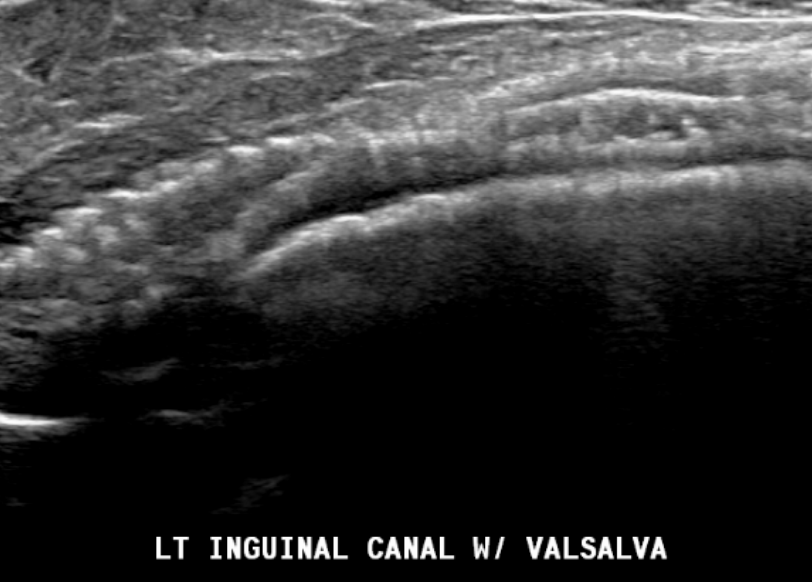
Mesh seen in the inguinal canal
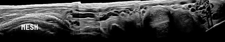
Mesh in inguinal canal along with vericoceles
Yonella